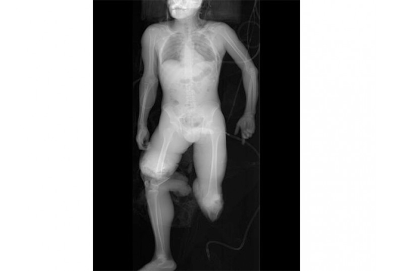
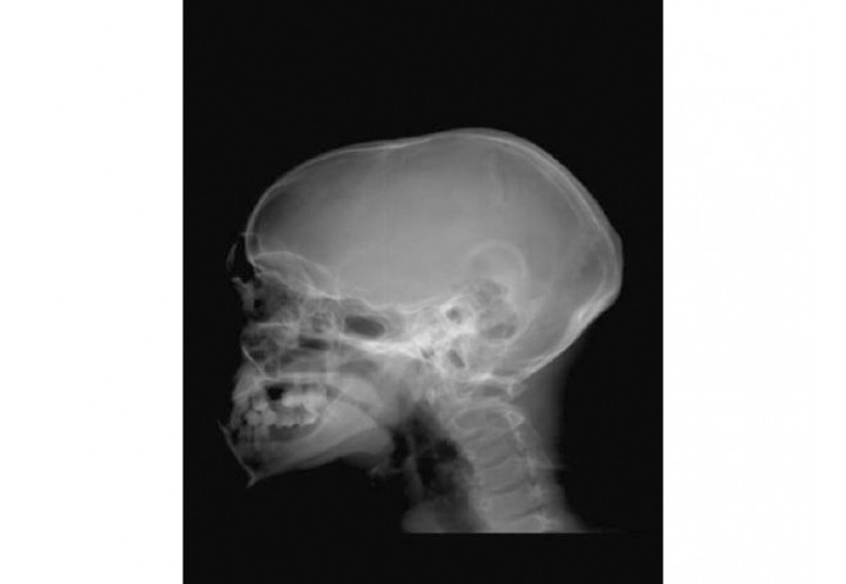
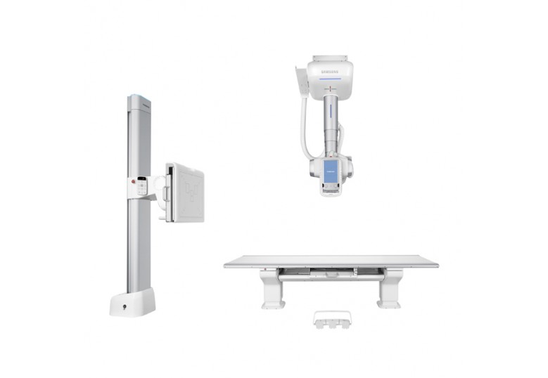
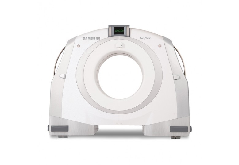
The Lodox plays a significant role in the initial management of trauma patients and is an important advance in the trauma imaging repertoire. The remarkable detection rate for injuries, low radiation dose and speed at which the whole body can be evaluated are advantages in the primary survey of acute trauma patients. Lodox provides time-saving, low dose investigation for emergency units with minimal interference in initial resuscitation.
Lodox provides a single (non-stitched), high-resolution radiographic image of the entire body. Lodox visualise skeletal, chest and pelvic pathologies ‘all-in-on’, and more accurately than conventional X-ray, in the primary trauma survey. Full-body imaging allows a better understanding of the patient’s entire injury pattern.
High Speed
A full-body trauma imaging study in two planes can be performed on the Lodox X-ray machine in 3-6 minutes. Rapid acquisition of radiographic detail is particularly important in ATLS resuscitation, where time predicts outcomes.
Low Radiation
Radiation emission and scatter are significantly lower than for conventional X-ray equipment. Together, these features improve safety for staff, significantly reduce radiation dose to patients, and allow uninterrupted resuscitation during imaging. Significantly lower radiation dose and high diagnostic image quality make Lodox a first-choice for paediatric poly-trauma.
Image Quality
Lodox high-definition, high-contrast images have been found to be better than or equal to conventional X-ray images for the detection of thoracic, pulmonary, mediastinal, pelvic and peripheral injuries. The unique, focused fan-beam of the linear slot-scanning technology improves image quality by reducing patient scatter image degradation, especially in larger patients.
Application
- Poly-trauma
- Gunshot wounds
- Foreign body ingestion
- Skeletal surgery
- Urinary stones
- Mass disasters
- Ventriculoperitoneal shunts
- Pediatric imaging
- Bariatric imaging
- Trauma in military medicine
Specifications
- Weight: 1500 kg / 3306 lbs
- Dimensions: 2810mm x 2276mm x 2250mm (LWH)
- Room height requirements: 2450mm
- Operating envelope: 2834mm x 2322mm x 2322mm (LWH)
- Required minimum area: 3798mm x 2808mm (LW)
Image Quality
- Contrast resolution: >16,000 grey levels (14 bits) - After log compression
Scanner throughput
- Linear scanning rate or speed (3 settings): 35 mm/s, 70 mm/s, 140 mm/s
- Beam width (FWHM @ 1000mm from focal spot): 1.4 - 2.8 mm
- Instantaneous frame rate (X-ray exposure duration at any one point): 11 - 80 milliseconds
- Time to complete a full scan: <13 seconds (nominally 12.98s at normal speed)
- Time from "end-of-scan" until a diagnostic image becomes available on the DVS screen: < 15 seconds (normal resolution image on a stand-alone 100 Mbits/s ethernet base-T network)
- Best case time between two successive X-rays on the same patient: 28 seconds (provided heat capacity of X-ray tube < 20%)
Output
- Radiation type: this equipment emits ionising radiation through an adjustable width, narrow slit, thereby producing fan-beam scanning across the patient. This slit is set to 0.4 mm (typically) or 1 mm (large patients)


